Elizabeth Hillman
Personalized Medicine
Neuroscience Meets Engineering
Elizabeth Hillman is developing cutting-edge research technologies to capture live images of the living brain using light to determine how the brain works and, in particular, how it regulates its blood flow.
Hillman’s fascination with medical imaging stems from childhood; she grew up doing gymnastics and experienced a number of injuries that led to doctor exams, medical imaging, and physical therapy sessions. The experience of having an MRI of her back during her teens and seeing herself “from the inside out” left her captivated.
X-Ray Visionary
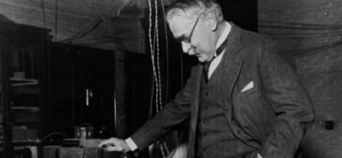
Michael I. Pupin
Alumnus and Former Professor
Michael I. Pupin (1858–1935), an 1883 Columbia College alumnus who returned to Columbia after graduation, served on the faculty for more than 40 years. During his tenure at the University, he taught mathematical physics and was one of the first faculty members to comprise the Engineering School’s newly created Department of Electrical Engineering. He taught many of Columbia Engineering’s great inventors, including Edwin Howard Armstrong (Class of 1913), maker of FM radio, and Irving Langmuir (Class of 1903), the pioneering surface chemist who was awarded the Nobel in 1932.
Pupin’s pioneering experiments with X-rays and fluorescent screens made it possible to produce X-ray images after only a few seconds. His 1896 discovery came on the heels of Wilhelm Roentgen, who is credited with developing X-rays that produced an image after several hours of exposure. The first use of the Pupin method, which utilized a fluorescent sheet supplied by Thomas Edison, was medical: he helped a surgeon determine where buckshot was imbedded in a patient’s hand.
Pupin received 34 patents for his inventions and won the Pulitzer Prize in 1924 for his autobiography, From Immigrant to Inventor, which tells the inspiring story of his upbringing in rural Serbia to his stature as one of the greatest American scientists of the early twentieth century. In addition to rapid X-ray imaging, he developed sonar technology and made revolutionary improvements to telegraphy and long-distance telephony. He was elected to the National Academy of Sciences in 1904.
At the Engineering School, Pupin helped lay the foundation for the Electrical Engineering Department, which continues to thrive and produce pioneering research and outstanding future engineers. Current faculty, researchers, and students have made important contributions in core areas, from communications and networking, signal processing, and digital and analog integrated circuits to electromagnetics and plasma physics, and photonics. In 1958, to mark the centenary of Pupin’s birth, the School established the Pupin Medal, given periodically to recognize service to the nation in engineering, science, or technology.
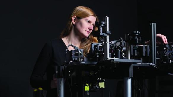
Elizabeth Hillman develops advanced in-vivo microscopy tools to study the living brain. She describes optical components as “very expensive Lego,” which when put together, properly make breathtaking images.
“Around this time, I discovered there was this thing called medical physics,” says Hillman, associate professor of biomedical engineering. “This was the perfect combination … using my skills in engineering and physics to have an influence on medicine. Pursuing my fascination with imaging and image formation is what got me here.”
By “here,” Hillman means concentrating on building new tools for brain imaging and performing neurovascularcoupling research in her lab. Hillman, who also has a joint appointment in Radiology, has received a multitude of honors and awards for her work in neuroscience and optics, and her investigations into neurovascular coupling—the relationship between local neural activity and subsequent changes in cerebral blood flow—are creating a lot of positive noise in research and medical communities.
“I’ve been developing my imaging and microscopy tools to let me study that interrelationship between neural activity and blood flow, trying to understand the mechanisms that govern the brain’s blood flow. This is really hard to study in the intact brain, so we need a lot of engineering to help us see deeper, faster, and at higher resolution,” she explains. “Quite recently we happened upon a new hypothesis that we think is a major component of blood flow regulation in the brain. Neuroscience over the years has focused so intently on neurons, they have barely noticed how important the blood vessels are.”
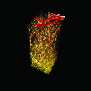
3D rendering of astrocytes and blood vessels in the living brain, imaged using Hillman’s home-built in-vivo two-photon microscopy system.
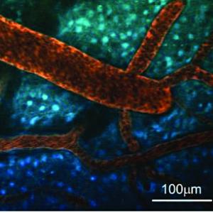
Astrocytes (blue) and blood vessels (red) in the living brain; Hillman studies the way that cells in the brain interact with blood vessels to regulate local blood flow.
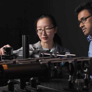
Students Thilanka Galwaduge (right) and Sharon Kim align their high-speed, dualbeam two-photon microscope for in-vivo brain imaging.
She explains that her work defining these new cellular mechanisms coupling neuronal activity to blood flow modulation, along with recent results exploring development of the newborn brain, and imaging the brains of patients during brain surgery has revealed how important regulation of blood flow is for brain health.
“Almost nobody has looked at the fact that the brain needs a lot of energy and considered how management of this energy could influence brain function and long-term brain health. Our research suggests there may be ways to look at brain disorders that go beyond looking at neurons. We are starting to look at Alzheimer’s disease, dementia, and even developmental disorders in a new way,” Hillman says.
Hillman has been interested in brain blood flow, and imaging it, since she was an undergraduate at University College London. Her senior project involved using near-infrared spectroscopy to measure changes in blood flow in her own brain. During her PhD studies, she developed a more complex 3D optical imaging system to measure blood oxygenation in the brains of premature babies.
Hillman says that her recent return to studying early brain development was partially influenced by watching the development of her two young sons. She was surprised to find so few brain imaging studies performed on children under the age of five.
“As a mother, I can completely understand how challenging it is to get young children to participate in research studies,” Hillman says. “However, I am just so in awe of the intense development that the brain goes through in these early years; it’s the period in which the brain learns basically every single thing that it does.”
As a result, Hillman recently started a project to develop new child-friendly brain imaging technologies that might make it possible to capture information about brain function and development while children play. Her goal is to better understand the brain during this critical period when autism, ADHD, and other developmental disorders are emerging, in the hope of providing new insights into diagnostics, monitoring therapy, and perhaps even the underlying basis of these and other conditions.
Hillman expects that her discoveries will soon start to change the way that doctors think about brain disorders and treating them in humans.
Almost nobody has looked at the fact that the brain needs a lot of energy and considered how management of this energy could influence brain function and long-term brain health. We are starting to look at Alzheimer’s disease, dementia, and even developmental disorders in a new way.