The Future. We're Working On It.
Jun 12 2017
Scientists attempting to decode the brain rely on high-speed recordings of individual neurons. Yet measuring enough neurons to understand how the brain drives behavior has remained a holy grail of neuroscience research.
Enter SCAPE, or swept confocally aligned planar excitation microscopy, a technique developed in the laboratory of Elizabeth Hillman, associate professor of biomedical engineering at Columbia Engineering. SCAPE enables 3D imaging of living tissues at very high speeds: 10–100 times faster than most in vivo microscopes. By imaging in 3D at such high speeds, SCAPE has the potential to transform neuroscience and biomedical research.
Another benefit of SCAPE is its simplicity. The technique requires no special preparation of samples and doesn’t require the user to move the sample or the microscope’s objective lens. As a result, SCAPE can capture real-time events in living animals, like the firing of neurons in a mouse brain or the beating of a zebrafish’s heart. SCAPE could also have clinical applications, providing doctors with a real-time 3D view of cells within a patient, for example, to guide removal of tumors.
Hillman, who is also associate professor of radiology at Columbia University Medical Center and a principal investigator at Columbia’s Mortimer B. Zuckerman Mind Brain Behavior Institute, first described SCAPE in a paper in Nature Photonics last year. She was subsequently awarded a $1.83 million, three-year grant from the NIH BRAIN (Brain Research through Advancing Innovative Neurotechnologies) Initiative to support her work on SCAPE.
Recognizing the significant commercial potential of SCAPE microscopy, in August 2016 Columbia entered into an exclusive, worldwide agreement with a leading microscope manufacturer to license SCAPE patents. The company plans to develop a commercial system, bringing the powerful technology to scientific laboratories around the world.
“We have received so much interest in SCAPE,” says Hillman, “we knew we needed help to get the technology onto the worldwide market as soon as possible. We are very excited to be entering this next phase and could not be more happy with our new partnership.”
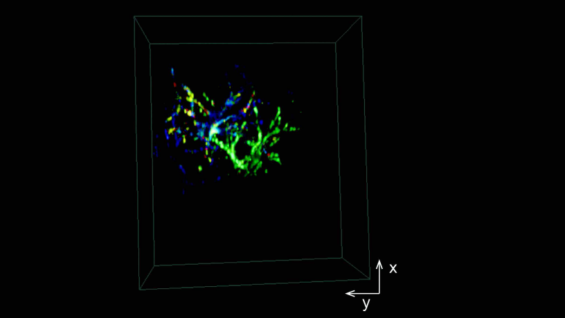
Apical dendrites of layer 5 excitatory neurons expressing calciumsensitive fluorophore GCaMP6 in an awake brain. SCAPE can capture spontaneous dendritic firing across a 600 micron square field of view to depths exceeding 250 microns at 10 volumes per second.